
Figure 1.
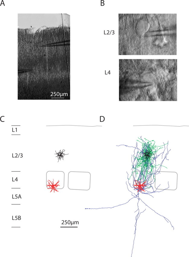
Paired whole-cell recordings of L4 spiny neurons and L2/3 interneurons. A, Bright-field image of the slice in the recording chamber after the experiment. B, DIC images at high magnification of the somata of the presynaptic and postsynaptic neuron. C, Computer reconstruction of presynaptic (red) and postsynaptic (black) dendritic geometries. Barrel outlines are superimposed. D, Reconstruction of dendrites and axons of the presynaptic (L4 spiny neuron, red and blue) and postsynaptic (L2/3 interneuron, black and green) neurons.