Figure 2.
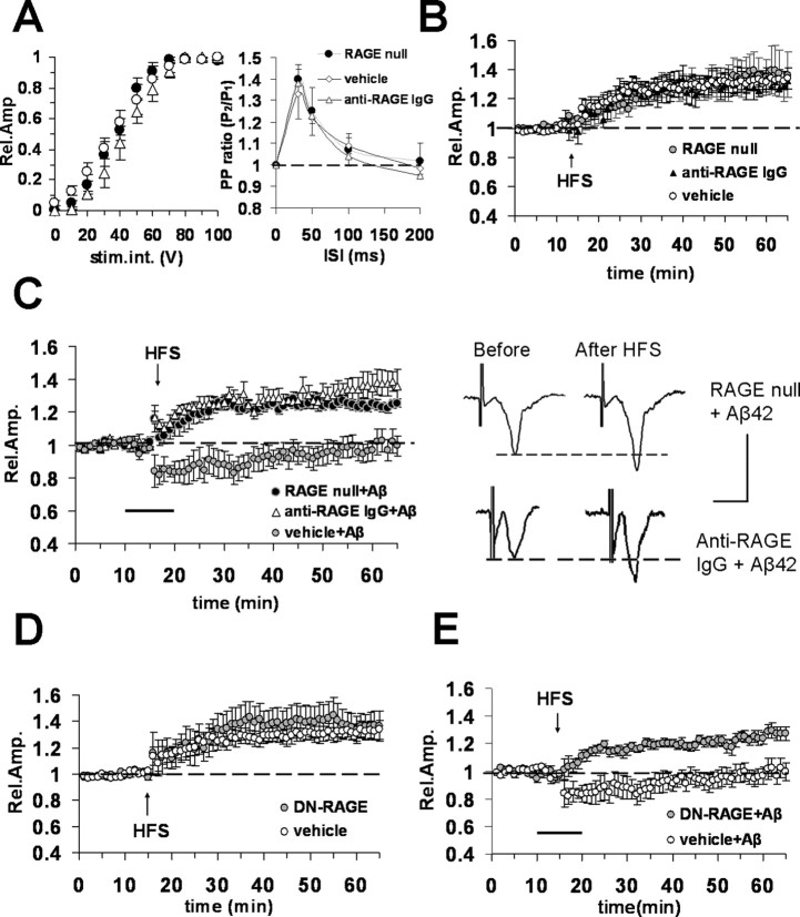
Blockade of RAGE protects against Aβ42 (200 nm)-induced reduction of LTP in entorhinal cortex slices. A, Left, Input–output curves did not show significant differences between vehicle-treated WT slices (open circles) and RAGE-null slices (filled circles) or WT slices treated with anti-RAGE IgG (open triangles). Right, Paired-pulse facilitation, PP ratio was not modified in RAGE-null slices and in slices from WT mice treated with anti-RAGE IgG with respect to vehicle-treated slices. B, LTP expression was unaffected in slices from RAGE-null mice (open circles) or in slices from WT mice preincubated with anti-RAGE IgG (2.5 μg/ml for 2 h, filled triangles) with respect to vehicle-treated slices (open circles). C, Left, Bath application (dark bar) of Aβ42 (200 nm) did not inhibit LTP in slices from RAGE-null mice (filled circles) or in slices from WT mice preincubated with anti-RAGE IgG (2.5 μg/ml for 2 h, open triangles) with respect to vehicle-treated slices (gray circles). Right, Representative field potentials recorded before and 50 min after HFS in RAGE-null and anti-RAGE IgG-treated slices from WT mice; bath application of Aβ was unable to block LTP. Calibration: 1 mV, 5 ms. D, LTP expression was comparable in vehicle-treated slices from WT mice (open circles) and in slices from Tg DN-RAGE (gray circles). E, Aβ42 (200 nm, bath applied, dark bar) failed to inhibit LTP in DN-RAGE slices (gray circles). Error bars indicate SEM.

