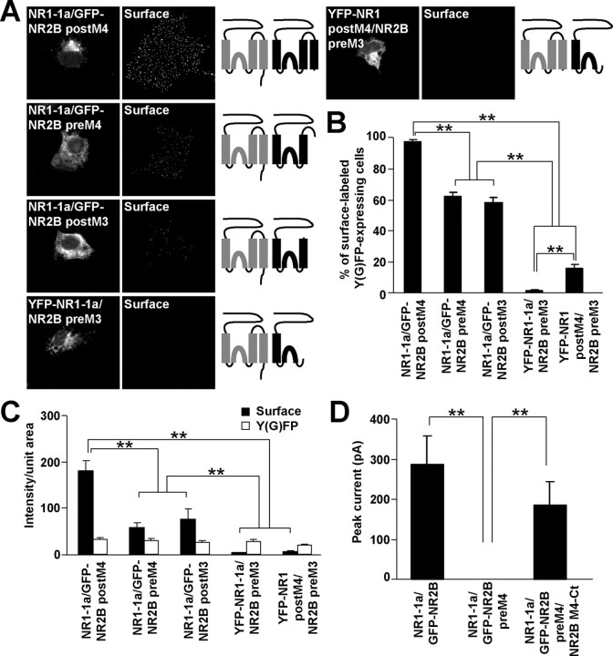
Figure 5.
NR2B M3 is a necessary structural component for negating the NR1 M3 ER retention signal. A, COS7 cells transfected with indicated combinations of cDNAs were stained for surface GFP. Schematic diagrams of membrane topology of NR1 (gray) and NR2B (black) subunits are shown. B, Bar graph represents the percentages of transfected COS7 cells exhibiting the surface staining in mean ± SEM; n >300 cells for each combination of cDNAs in three experiments. C, Quantification of surface (black) and total Y(G)FP (white) expression of indicated combinations of cDNAs. Data show mean ± SEM of fluorescence intensities of >45 transfected cells measured in three experiments. D, GFP-NR2B preM4 can form a functional channel after coexpression with NR2B M4-Ct and NR1-1a. Currents evoked by the application of 1 mm glutamate and 1 mm glycine were recorded from HEK293 cells expressing indicated combinations of cDNAs. The mean peak current amplitudes ± SEM are shown; n = 6. **p < 0.001, ANOVA.

