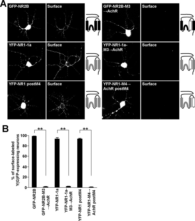
Figure 9.
Replacement of both NR2B and NR1 M3 for AchR α M4 and NR1 M4 for AchR α M3 inhibits surface targeting of the NMDA receptors in cortical neurons. A, Cortical neurons transfected with indicated combinations of cDNAs were immunostained for surface Y(G)FP. Schematic diagrams of membrane topology of NR2B (black) and NR1 (gray) subunits with replaced M3 or M4 for AchR α M (white) are shown. B, Bar graph represents the percentages of Y(G)FP-expressing cortical neurons exhibiting the surface staining in mean ± SEM; n >100 neurons in three experiments. **p < 0.001, unpaired t test.

