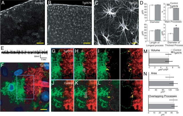
Figure 7.
Cortical astrocytes in seizure-free Tg2576 transgenic mice are reactive but do not lose their domain organization. A, Control cortex with low GFAP expression throughout cortex. Scale bar (SB), 100 μm. B, Cortex of a Tg2576 mouse with upregulation of GFAP throughout all cortical layers. SB, 100 μm. C, Higher-power image of cortical astrocytes in Tg2576 mouse. SB, 10 μm. D, Morphometric analysis based on GFAP. Astrocytes in the Tg2576 mice exhibited only a significant increase in the maximum thickness of GFAP-positive processes (Tg2576, n = 65, 3 mice; mean ± SEM; *p < 0.001). E, Representative EEG recording of Tg2576 mice. F, Diolistic labeling of cortical astrocytes in Tg2576 mouse. SB, 10 μm. G–I, High-power image of blue box in F showing limited overlap between processes of two adjacent astrocytes. J–L, Adjacent control astrocytes showing limited overlap and extension of processes into the adjacent cell's domains. M, Quantification of the volume of reactive astrocytes in Tg2576 compared with control. There is no significant increase in the volume of reactive astrocytes in the Tg2576 model (n = 12, 3 mice; mean ± SEM; p = 0.45). N, Quantification of amount of overlap between adjacent cells in Tg2576 and control mice (Tg2576, n = 30, 3 mice; mean ± SEM; there is no significant increase in amount of overlap compared with control; area, p = 0.5; overlapping process, p = 0.8).

