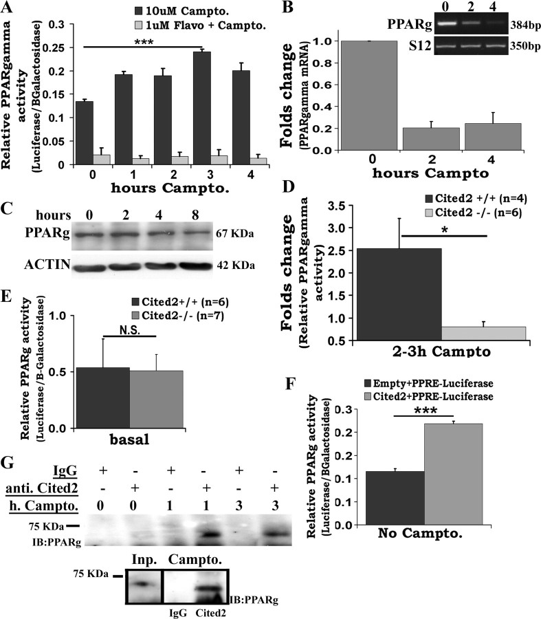
Figure 8.
PPARγ activity, but not levels, is upregulated after DNA damage in a Cdk-Cited2-dependent manner. A, PPARγ activity increases after DNA damage and is blocked by the Cdk inhibitor flavopiridol. Seventy-two hours after plating, cortical neurons were transiently cotransfected with PPRE-luciferase- and CMV-β-galactosidase-expressing plasmids using Lipofectamine as described in Materials and Methods. Twenty-four hours after, cells were treated with 10 μm campto and/or 1 μm flavopiridol for specified times. Data represent values of luciferase/β-galactosidase activity. Bars represent the mean ± SEM from four independent experiments. ***p < 0.001. B, PPARγ message decreases after DNA damage. Cortical neurons were treated with campto at indicated times. Total RNA was extracted and RT-PCR result from one representative experiment is shown in inset. Normalized densitometry data are presented as fold change relative to nontreated sample. Each bar represents the mean ± SEM from three independent experiments. C, Total protein was extracted from campto-treated cortical neurons and analyzed by Western blot as described. Results from one representative experiment are shown. D, Cited2 deficiency blocks PPARγ activity upregulation by campto. Seventy-two hours after plating, cortical neurons from littermate embryos from heterozygous Cited2 crosses independently cultured were transfected and treated with 10 μm campto to assess PPARγ activity as described above. Values represent fold change of normalized luciferase activity (luciferase/β-galactosidase) of correspondent sample relative to transfected nontreated sample. Each bar represents the mean ± SEM from n embryos. *p < 0.05. E, Basal PPARγ activity does not differ between Cited2 KO and WT neurons. PPARγ activity was measured as described in D and presented as normalized luciferase activity (luciferase/β-galactosidase). The bars represent the mean ± SEM from n independently cultured embryos. N.S., No significant difference. F, Cited2 overexpression induces PPARγ activity. Seventy-two to 96 h after plating, cortical neurons were transiently cotransfected with empty pcDNA3 or pcDNA3-Cited2-, PPRE-luciferase-, and CMV-β-galactosidase-expressing plasmids using Lipofectamine. Twenty-four hours after, luciferase activity was evaluated as described in Materials and Methods. Data are presented as normalized luciferase activity (luciferase/β-galactosidase). Each bar represents the mean ± SEM from three independent experiments. ***p < 0.001. G, Cited2 and PPARγ interact after DNA damage. Cortical neurons were treated with campto at indicated times and total protein was extracted as indicated in Materials and Methods. Control IgG or anti-Cited2 antibodies were incubated with total cell lysates (∼2 × 10⁷ neurons). Antibodies were isolated by IP beads and resolved by SDS-PAGE followed by anti-PPARγ Western blot. Results from two independent experiments are shown. Inp., Input, TCA precipitate from total cell lysate (∼107 untreated neurons).

