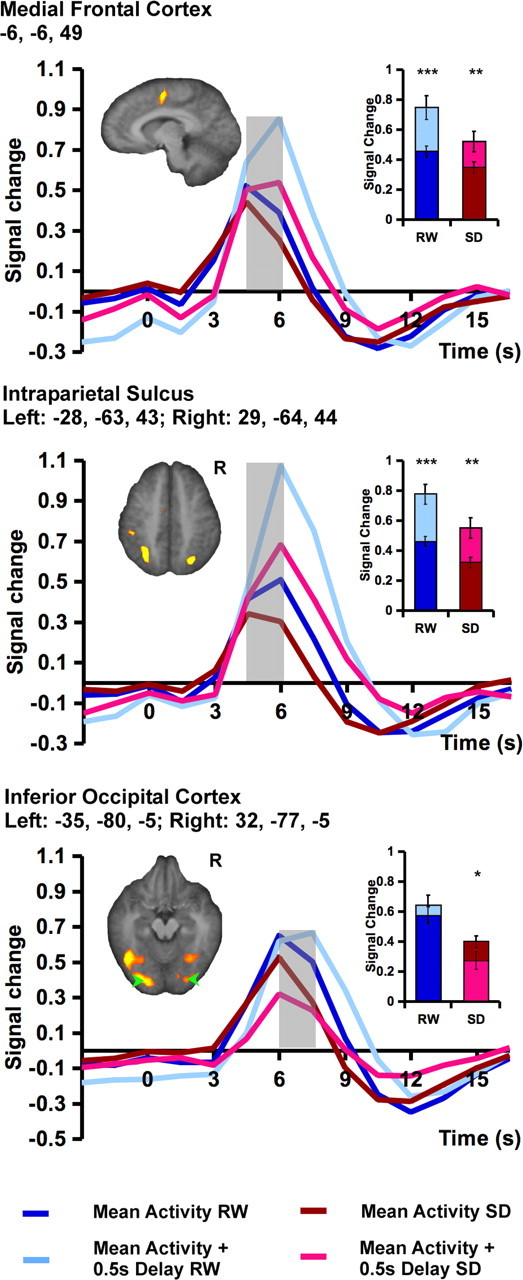
Figure 5.

Task-related fMRI signal associated with responding at the average RT and during a lapse (modeled as a response 0.5 s longer than the average RT for that state). Lapses were associated with higher peak signal in the medial frontal cortex (top) and bilateral intraparietal sulcus (middle) in both RW and SD. In the occipital region, lapsing significantly reduced peak signal during SD (bottom) but did not significantly modulate the peak fMRI signal after RW, although there was a delay in the time-to-peak. Random effects analysis using a threshold of p < 0.001 was used to detect task-related activation. Significant differences between peak signal associated with a lapse trial and the mean response for each state are marked with an asterisk. The shaded time points indicate those contrasted to assess significant state effects. The inset shows the mean peak signal associated with the time points under consideration. Error bars represent SEM. *p < 0.05, **p < 0.005, ***p < 0.001.
