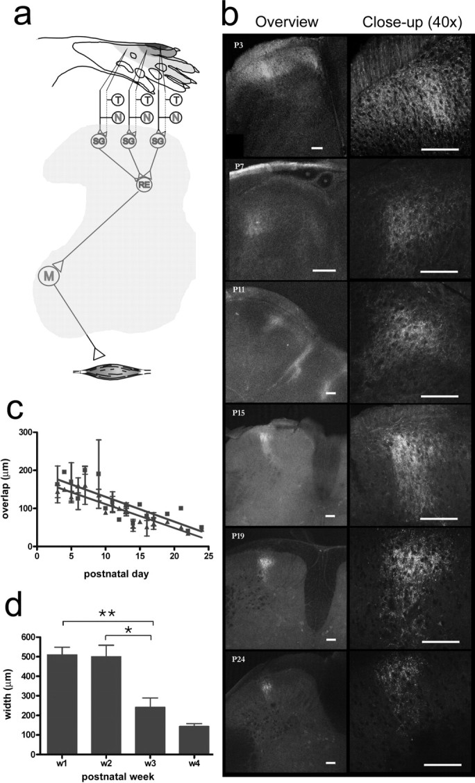
Figure 4.

Developmental reorganization of coarse and thin afferent terminations from the NWR receptive field of the PL. a, Schematic of the immature PL NWR module: note the tactile-nociceptive convergence in the superficial dorsal horn. T/N, Tactile/nociceptive; SG, substantia gelatinosa interneurons; RE, reflex encoding multimodal interneurons; M, motor neurons. The quantified PL NWR receptive field (top) is shown as blue shaded isoresponse areas; colors denote >70%, 30–70%, and 0–30% of maximum values. b, Examples of images collected at different developmental time points (P3–P24). CTb-labeled fibers (red) traverse the WGA (green)-labeled zone in the first 2 weeks. During the third week, the laminar overlap decreases, and terminal fields narrow mediolaterally into a columnar structure. c, The mediolateral (red) and dorsoventral (green) maximum width of the overlap zone of thin and coarse afferents (postnatal days 3–24; linear regression slopes/0; p < 0.0001). d, Mediolateral maximum width of the thin fiber terminal field during postnatal development (p < 0.0001, Kruskal–Wallis test, *p < 0.05; **p < 0.01, Dunn's multiple-comparison post test. Scale bars, 100 μm.
