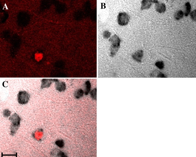
Figure 1.
Three confocal images of the same brain section demonstrating double-labeling of Hu (brown) and BrdU (florescence red). Only one (red nucleus) of the several neurons (Hu+, brown) shown in this field was born at the time of BrdU injections. A, BrdU labeling. B, Hu labeling. C, BrdU and Hu labeling. Scale bar, 10 μm.

