
Figure 1.
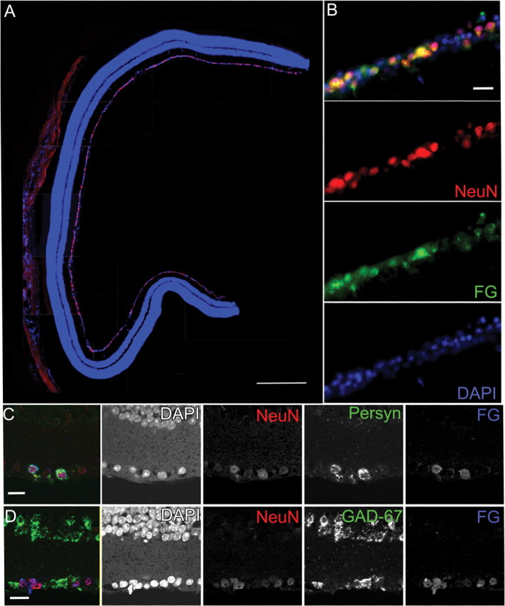
Immunohistochemistry and in situ hybridization of RGCs in the DBA/2 retina showing that NeuN antibody predominantly labels RGCs. A, A vertical section through a 3-month-old DBA/2 retina immunolabeled with NeuN (red) and the nuclear stain DAPI (blue). Although occasionally observed in the inner nuclear layer, the majority of NeuN-positive cells are observed in the GCL. Scale bar, 250 μm. B, Higher-magnification view of double immunolabeling in the GCL for NeuN (red) and FG (green) with DAPI (blue) shows that all retrogradely labeled FluoroGold-positive RGCs are also NeuN positive. The panels broken out by fluorescence color channel allow one to visualize the variations in labeling intensity for both NeuN and FG. Scale bar, 50 μm. C, In situ hybridization combined with immunohistochemistry permitted colocalization of NeuN immunolabel, Sncg mRNA, and FG immunolabel in the GCL of a 3-month-old DBA/2 retina. All FG-positive RGCs were also NeuN positive and had Sncg mRNA. Breakout panels show the individual fluorescence color channels. Scale bar, 50 μm. D, In situ hybridization for the amacrine marker GAD-67 combined with immunohistochemistry for NeuN and FG show that all FG-positive cells are NeuN positive; FG-positive/NeuN-positive cells are GAD-67 negative. The individual fluorescence channels allow one to discern light NeuN immunolabeling in a small number of GAD-67 displaced amacrine cells. Scale bar, 50 μm.