
Figure 3.
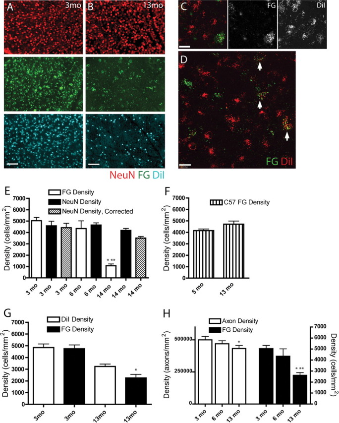
RGC axon pathology assessed by comparing two retrograde labeling methods. A, Separate fields from 3 month (3mo) whole-mount retina immunolabeled with NeuN (red) and retrogradely labeled with FG (green) and DiI (blue). NeuN, FG, and DiI showed similar numbers across all retinas at 3 months. Scale bar, 100 μm. B, By 13 months (13mo), FG labeling (middle panel, green) in all retinas was dramatically reduced, and DiI labeling (bottom panel, blue) was reduced to a lesser extent, whereas NeuN labeling was unaffected (top panel, red). Immunohistochemistry for FG was done to improve the tracer signal; puncta smaller than somal size in the FG panel are nonspecific secondary antibodies within the tissue. Scale bar, 100 μm. C, High-magnification image of typical whole-mount retinal field of RGCs retrogradely labeled with FG (green) and DiI (red) at 14 months showing more DiI-positive than FG-positive cells. The cell in the lower right is FG and DiI positive. Scale bar, 20 μm. D, High-magnification field of DiI-FG retrogradely labeled DBA/2 retina whole-mount. Arrows point to RGCs with varying levels of FG and DiI labeling. The majority of cells are DiI positive. Scale bar, 20 μm. E, Graph of RGC density in DBA/2 retinal whole mounts as quantified with NeuN immunolabel and FG tracer. Data from 3 month (3 mo) and 14 month (14 mo) retinas are presented with (hash marked) and without (black) the displaced amacrine cell correction factor. No correction factor was available for 6 month (6 mo) DBA/2 data. NeuN and FG labeled a similar number of RGCs at 3 and 6 months, but at 14 months, FG labeling was markedly reduced compared with earlier time points (*) and compared with NeuN immunolabel in 14 month retinas (**) (p < 0.01). F, Graph of FG-labeled RGC density in retinal whole mounts of 5 month (5 mo) and 13 month (13 mo) C57BL/6 normal controls. Unlike DBA/2 retinas, there was no reduction in FG-labeled RGCs in older C57BL/6 controls. G, Graph of RGC density in DBA/2 retinal whole mounts labeled with both FG and DiI. FG and DiI labeling was similar at 3 months (3mo). By 13 months (13mo), there was a visible but not statistically significant decrease in DiI-positive RGCs, whereas the FG-positive (*) RGCs were significantly reduced (p < 0.05). H, Counts of RGC axons in the optic nerve from FG-quantified retinas showed a significant decrease by 13 months (*) (p < 0.05). FG retrograde labeling of RGCs at 13 months was significantly decreased relative to FG labeling at both 3 (*) and 6 (**) months (p < 0.05). As a percentage of the total, FG loss in the DBA/2 retinas was larger than axon loss in the corresponding optic nerves.