
Figure 1.
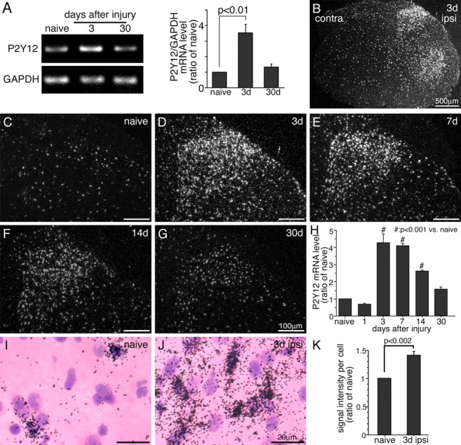
PSNL increases P2Y12 expression in the ipsilateral spinal cord. A, The levels of P2Y12 mRNA in the ipsilateral L4–L5 spinal cord were determined using the RT-PCR technique. Gel panels show PCR products from the L4–L5 spinal cord taken from naive and 3 and 30 d after surgery. Right graph shows quantification of the relative mRNA levels of P2Y12 in the spinal cord. P2Y12 mRNA levels were normalized against corresponding control (mean ± SEM) (p < 0.01 compared with naive). B–G, Dark-field images of ISHH show P2Y12 mRNA in the spinal cord of a naive rat (C), 3 (B, D), 7 (E), 14 (F), and 30 (G) days after PSNL. Scale bars: B, 500 μm; C–G, 100 μm. D–G show the dorsal horn ipsilateral to the surgery. H, Quantification of the silver grains in dorsal horn (laminas I–III) (n = 4, 10 sections at each time point). P2Y12 signals were normalized against naive rat. # p < 0.01 compared with naive control. I, J, Higher-magnification photographs of laminas II–III of the dorsal horn under bright-field illumination in the naive rat (I) and 3 d after PSNL (J). Scale bars, 20 μm. K, The mean intensity of P2Y12 mRNA signals per labeled cells in ISHH tissue sections of naive rats and 3 d after the surgery. p < 0.002 compared with naive control. GAPDH, Glyceraldehyde-3-phosphate dehydrogenase; contra, contralateral; ipsi, ipsilateral.