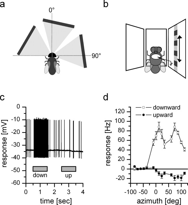
Figure 2.
Intracellular recording from DNOVS2. Top view (a) and frontal schematic drawing (b) of the stimulus situation. c, Example response of DNOVS2 to full field downward and upward motion in all three monitors. The cell responds to downward motion with an increase of the spike frequency and to upward motion with a slight decrease of the spike frequency. d, Response of DNOVS2 to vertical motion as a function of the azimuth position. The highest responses to vertical motion are elicited at two positions of the azimuth: at a frontal position (10°) and a lateral position (75°) with downward motion increasing the spike rate and upward motion decreasing the spike rate. The mean firing rate at rest is 5–15 Hz and is increased by 88 Hz at the positions with highest response. In between, the response is less with a local minimum at 35° and an increase of the firing rate by 55 Hz. Data represent the mean ± SEM recorded from n = 7 flies.

