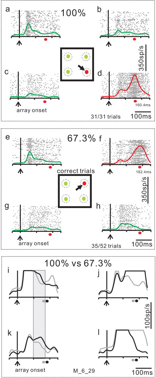
Figure 4.

Example of target and distractor neuronal activity in the selection task. An example set of four neurons recorded simultaneously during performance of the selection task. a–c, SC neuronal activity recorded in the target selection task when distractors were located in the RF of the recorded neuron (distractor neurons). d, SC neuronal activity when the target was located in the neuron's RF (target neuron). In each panel, each tick is an action potential and each row of ticks is one trial. The envelope of activity (SDF, σ = 10 ms) is superimposed on the rasters. Red SDFs are from target neurons and green SDFs are from the distractor neurons. Each of the four panels is aligned on the onset of the array of spots indicated by the vertical line and the upward arrow (array onset). The square in the center of the four panels show the stimulus display. The black arrow indicates the correct choice of saccade to the differently colored target. The dashed circles are schematics of neuronal RFs to emphasize the fact that we recorded from four neurons simultaneously. The trials shown in this figure are sorted based on performance. a–d show the neuronal activity when the monkey accurately performed all trials with the target located at the 315° position (31 of 31 trials, 100%). The filled red circle below each panel marks the average saccade latency made to the correct target for reference. e–h show the subset of trials in which the monkey chose the saccade target correctly when it appeared at the 45° position. The monkey performed the 45° position trials less well (35 of 52 trials; 67.3% correct) compared with the 315° position trials. i–l directly compare the SDFs for the trials in which the monkey performed the 315° target with 100% accuracy and the trials in which the monkey performed the 455° target with 67.3% accuracy. The gray traces are the same as those shown in a–d. The black traces are the same as those shown in e–h. Note the expanded vertical scale. The gray filled circle on the abscissa is the saccade latency on the 100% correct trials. The black filled circle on the abscissa is the saccade latency on the 67.3% correct trials. The shaded great rectangle shows that the distractor neuronal activity was different in these two subsets of trials. The M_6_29 indicates the data come from monkey m and the date, 6_29 was used as a file identifier.
