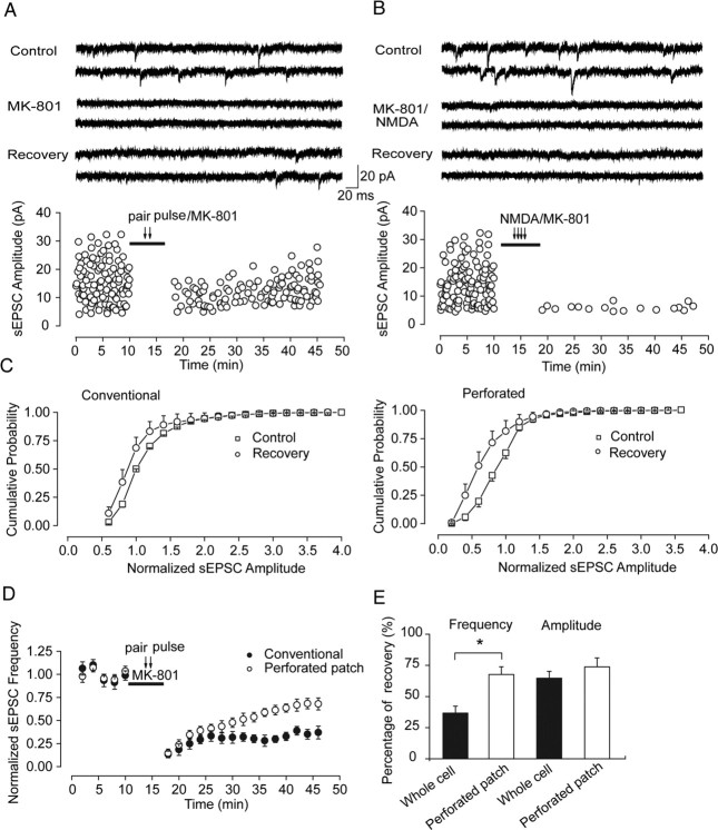
Figure 3.
Increased recovery level of NMDAR-mediated spontaneous EPSCs in perforated patch mode suggests the possible involvement of intracellular components in recovery. A, Selective block of synaptic NMDAR-mediated spontaneous EPSCs recorded in conventional whole-cell patch mode. Top, Representative traces from neurons at the indicated experiment conditions (control, paired-pulse stimulation/MK801, recovery). Bottom, Spontaneous NMDAR EPSCs recorded at different time points before and after selective synaptic NMDARs block through paired-pulse stimuli given at 0.125 Hz accompanied by MK-801 application. After 30 min MK-801 washout, partial recovery of EPSCs was observed. B, Agonist-evoked block of NMDAR-mediated spontaneous EPSCs recorded in conventional whole-cell patch mode. Top, Representative traces from neurons at the indicated experiment conditions (control, NMDA/MK801, recovery). Bottom, Complete and irreversible block of spontaneous NMDAR current after whole-cell coapplication of agonist NMDA and MK-801. C, Cumulative distribution of sEPSC amplitude (mean ± SEM) before MK-801 application (control) and after recovery from MK-801 (recovery) recorded in conventional whole-cell patch mode (left) and perforated patch mode (right), respectively. D, Partial recovery of NMDAR-mediated sEPSC frequency in two recording modes. Normalized sEPSC frequency over different time points show significantly increased recovery level in perforated patch mode than that in conventional patch mode. E, Statistical graphs show partial recovery of sEPSC amplitude and frequency in two recording modes, with frequency recovered up to 36.65 ± 5.63% of the initial level in conventional patch mode compared with 67.55 ± 6.22% in perforated patch mode (*p < 0.05). Amplitude recovery was 65.12 ± 6.46% in conventional patch mode compared with 73.85 ± 7.13% in perforated patch mode (no significance).

