
Figure 1.
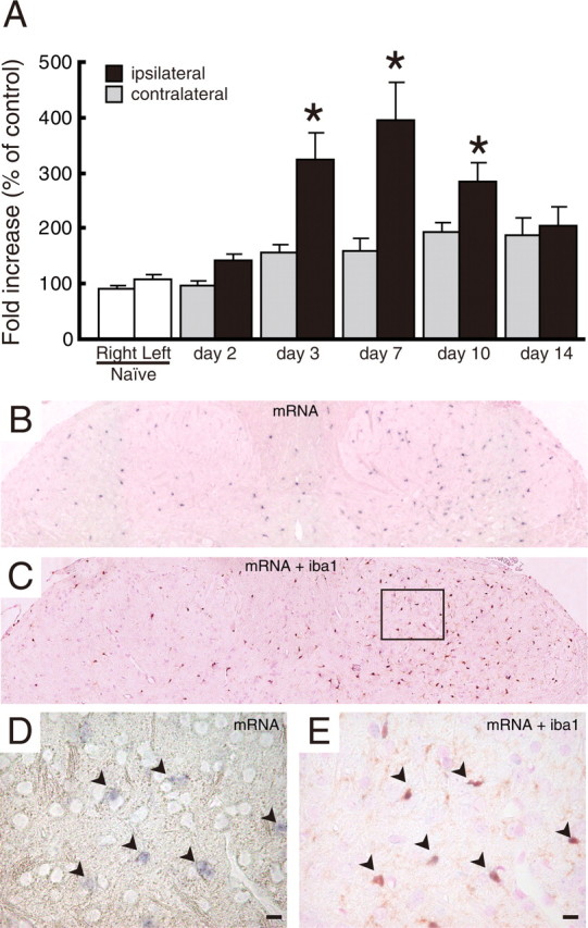
Upregulation of P2Y12R mRNA after peripheral nerve injury. Quantitative and histological analyses of P2Y12R mRNA expression in the spinal cord are shown. A, Total RNA extracted from rat spinal cord was subjected to quantitative analysis of P2Y12 mRNA expression after peripheral nerve injury by real-time PCR. Bar graphs show the average fold increase in the level of P2Y12 mRNA expression in spinal cord hemisections compared with the mean expression level of P2Y12 mRNA in naive animals. Each measurement was normalized to GAPDH content. Data are means ± SEM of five individual animals (*p < 0.05 vs the naive spinal cord, one-way ANOVA post hoc Tukey's test). B, A DIG-labeled RNA probe specific for P2Y12 mRNA was visualized by in situ hybridization in rat spinal cord, 7 d after the nerve injury. C, An RNA-probed slice was subsequently stained with anti-Iba1 antibody and visualized with DAB staining. D, E, Magnifications of the squared area show the sections before and after Iba1 staining. Arrowheads indicate colocalization of P2Y12 mRNA-positive cells and Iba1 signals. Scale bars, 10 μm.