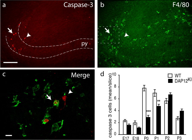
Figure 1.
Apoptotic neurons in neonate hippocampus are in contact with microglia. Double labeling of P0 hippocampus with antibodies against activated caspase-3 (red) (a) and F4/80 (green) (b). The arrow and arrowhead indicate caspase-3-positive cells that are apposed or not with microglia, respectively. Scale bar, 100 μm. py, Pyramidal cell layer. c, Higher magnification of the subiculum. Activated caspase-3-positive cells are surrounded (arrow) or contacted (arrowhead) by microglial processes. Scale bar, 10 μm. d, Quantification of developmental death in the hippocampus between E17 and P3 in WT (white bar) or in DAP12KI (black bar). Shown are mean ± SEM. ***p < 0.0001; **p < 0.001.

