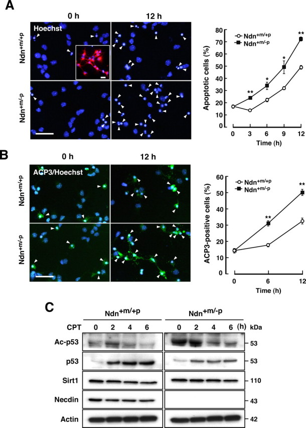
Figure 5.

Necdin deficiency enhances CPT-induced apoptosis of cortical neurons. A, Apoptosis of CPT-treated neurons. Primary neurons were prepared from the cerebral cortex of wild-type (Ndn+m/+p) and necdin-deficient (Ndn+m/−p) mouse forebrain at E14.5. Dissociated cell cultures were treated with CPT (10 μm) and stained with Hoechst 33342. Arrowheads indicate the apoptotic cells. Neuronal enrichment was evaluated by immunostaining MAP2 (inset). Scale bars, 20 μm. Neurons with condensed or fragmented nuclei were counted at the indicated times (>250 cells; mean ± SEM; n = 3) (graph). *p < 0.05; **p < 0.01. B, Neurons containing ACP3. Primary neurons were treated with CPT as in A and stained with an antibody to ACP3. Scale bar, 20 μm. ACP3-immunopositive cells were counted (>220 cells; n = 3) (graph). **p < 0.01. C, Western blot analysis. Neuronal lysates (7 μg per lane) were prepared at the indicated times of CPT treatment and analyzed by Western blotting with antibodies against acetyl-p53 (Lys373) (Ac-p53), p53, Sirt1, necdin, and actin.
