
Figure 6.
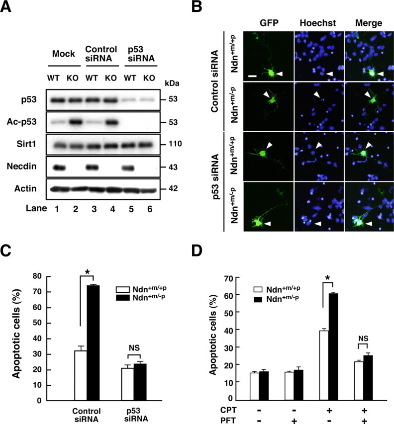
Necdin specially suppresses p53-dependent neuronal apoptosis. A, Western blot analysis. Cortical neurons were prepared from wild-type (Ndn+m/+p, WT) and necdin-deficient (Ndn+m/−p, KO) mouse forebrain and transfected with p53 siRNA and negative control siRNA (Control siRNA). Neurons were harvested 24 h later, and the lysates were immunoblotted with antibodies to p53, acetyl-p53 (Lys373) (Ac-p53), Sirt1, necdin, and actin. B, Immunostaining. Cortical neurons transfected with negative control siRNA (Control siRNA), and p53 siRNA were visualized by cotransfected GFP and immunostained with Hoechst 33342 (arrowheads). Scale bar, 10 μm. C, Quantification of apoptotic neurons. Neurons with condensed or fragmented nuclei among >30 GFP-positive cells were counted (mean ± SEM; n = 3). D, Inhibition of p53-dependent apoptosis by pifithrin-α (PFT). Neurons were treated with PFT (200 nm) and CPT (10 μm) for 9 h and stained with Hoechst 33342. Neurons with condensed or fragmented nuclei were counted (>200 cells; mean ± SEM; n = 3). *p < 0.01. NS, Not significant (p > 0.05).