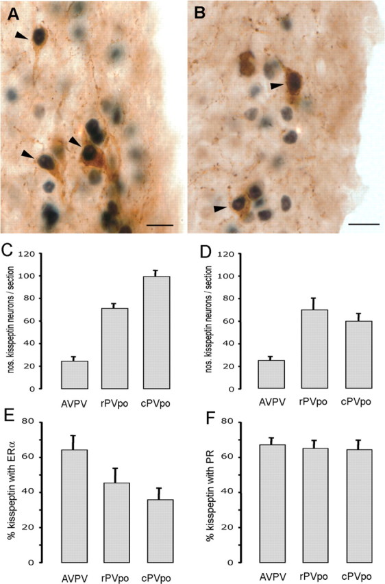
Figure 1.

RP3V kisspeptin neurons express ERα and PR. A, Dual-label immunocytochemistry showing AVPV kisspeptin neurons (brown) with ERα-immunoreactive nuclei (black). Arrowheads indicate dual-labeled cells. B, Dual-label immunocytochemistry showing PVpo kisspeptin neurons (brown) with PR-immunoreactive nuclei (black). C, Bar graph showing the mean + SEM number of kisspeptin-immunoreactive neurons counted per section at the three levels of the RP3V (AVPV, rPVpo, and cPVpo) in the ERα dual-labeling study. D, Bar graph showing the number of kisspeptin neurons counted per section in the AVPV, rPVpo, and cPVpo in the PR dual-labeling study. E, F, Percentage of kisspeptin neurons expressing ERα (E) and PR (F) throughout the RP3V. Scale bars, 10 μm.
