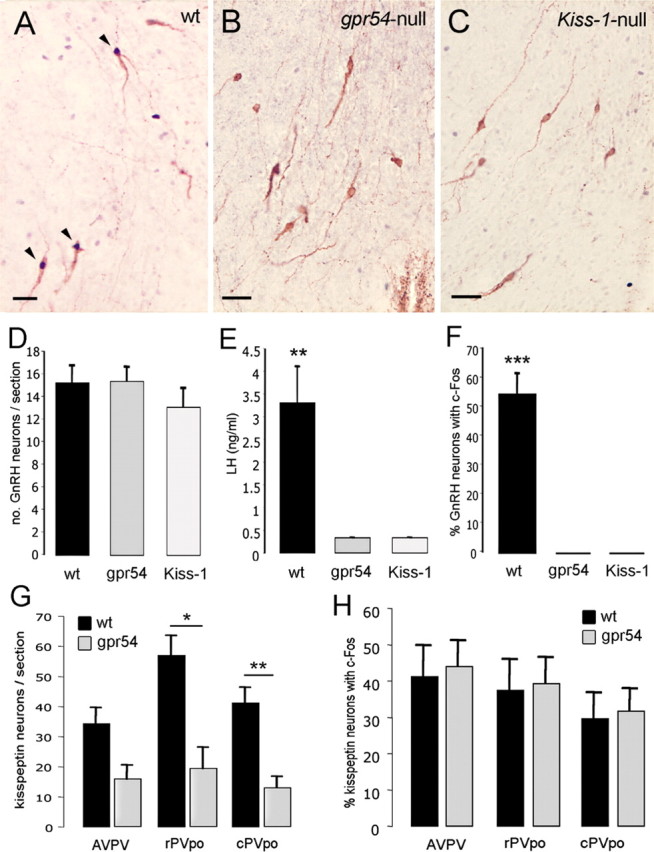
Figure 3.

Both the LH surge and GnRH neuron activation are absent in Gpr54- and Kiss1-null mice. A, Dual-label immunocytochemistry showing three GnRH neurons (brown) expressing c-FOS (black nuclei) in wild-type siblings. B, Dual-label immunocytochemistry showing absence of c-FOS staining in GnRH neurons of Gpr54-null mice. C, Dual-label immunocytochemistry showing absence of c-FOS staining in GnRH neurons of Kiss1-null mice. D, Bar graph showing the mean + SEM number of GnRH neurons counted per section in rPOA of wild-type siblings (wt) and Gpr54- (gpr54) and Kiss1- (Kiss-1) null mice. E, Mean + SEM LH levels in OVX+E+P mice of the three genotypes. F, Percentage of GnRH neurons expressing c-FOS in OVX+E+P mice of the three genotypes. G, Bar graph showing the mean + SEM number of kisspeptin neurons counted per section in the AVPV, rPVpo, and cPVpo of wild-type and Gpr54-null mice. H, Percentage of kisspeptin neurons expressing c-FOS in the AVPV, rPVpo, and cPVpo of OVX+E+P wild-type and Gpr54-null mice. *p < 0.05, **p < 0.01, ***p < 0.001, wt versus gpr54 or Kiss-1 or as indicated. Scale bars, 20 μm.
