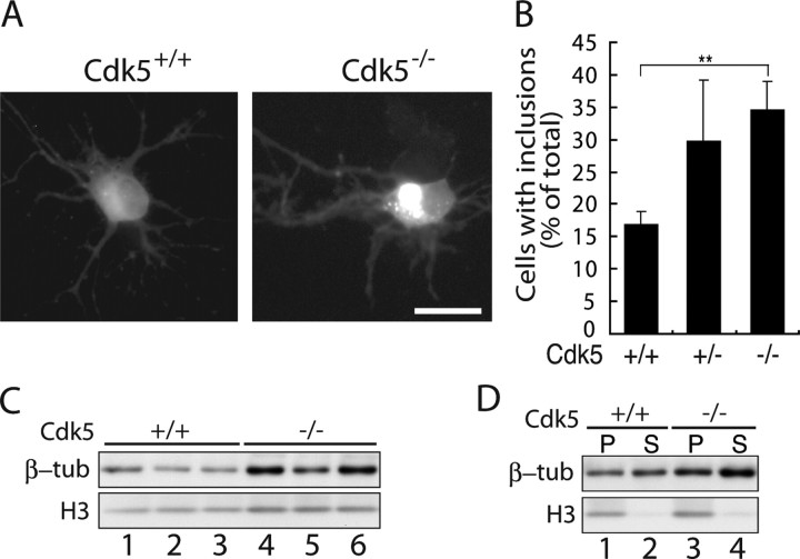
Figure 6.
mhtt inclusion formation in primary cultured neurons of Cdk5−/− mouse. A, Fluorescence images of EGFP in Cdk5+/+ or Cdk5−/− mouse cortical primary neurons transfected with tNhtt-166Q-EGFP. Scale bar, 10 μm. B, Neurons containing EGFP inclusions were counted and expressed as the percentage ratio to total EGFP-positive cells (mean ± SEM; **p < 0.001). C, Immunoblot showing the expression of β-tubulin in control and Cdk5−/− neurons. D, Immunoblot showing the amount of β-tubulin in the MT polymers (P) and protomeric soluble pool (S) in Cdk5−/− and control neurons.

