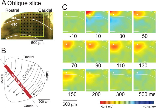
Figure 8.
The spread of activity in oblique sections (perpendicular to the optic tract). The slice was cut in the oblique direction to avoid an effect of optic fiber stimulation. A, Position of electrode on the slice. B, A schematic illustration of the cutting direction. C, The computed color images of field potentials evoked from a biphasic pulse to the electrodes marked with a white square. Representative images from n = 12 slices are shown.

