
Figure 9.
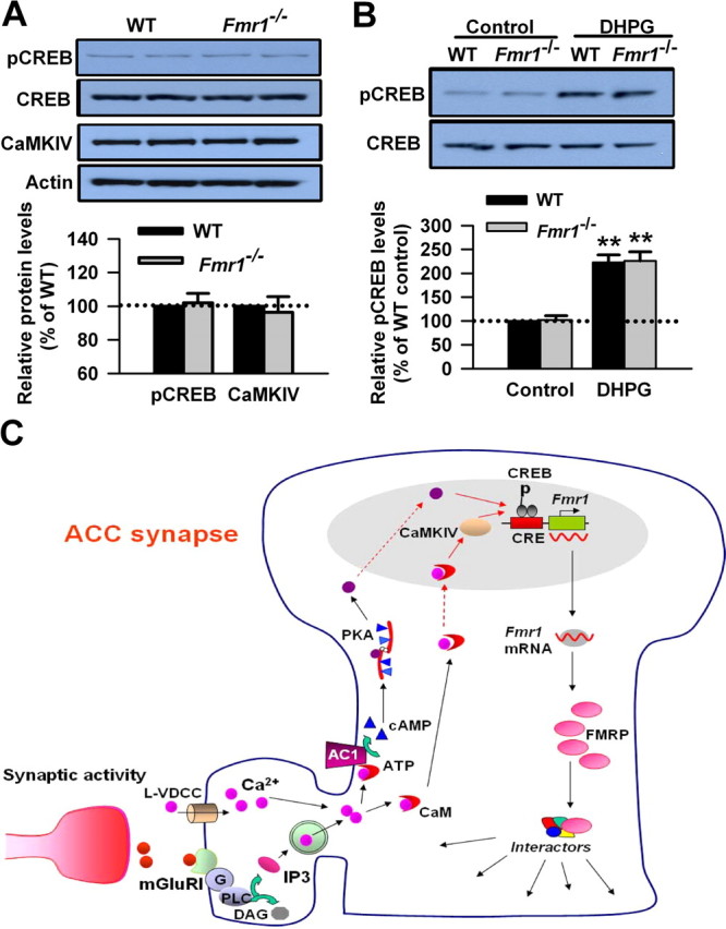
FMRP is downstream of the group I mGluR–CREB pathway in ACC neurons. A, There is no difference in basal levels of CREB in ACC between WT and Fmr1 KO mice. The basal levels of CaMKIV were not affected in ACC of Fmr1 KO mice compared with that of WT mice. B, The phosphorylation of CREB induced by DHPG treatment was not affected in Fmr1 KO mice compared with WT mice. The slices were treated with DHPG (100 μm) for 15 min. Representative Western blot (top) and quantification data (bottom) of pCREB levels are shown for corresponding treatments. Data were normalized by WT control values. **p < 0.01 compared with control; n = 4 mice for each group in A; n = 6 mice in B. C, The proposed signaling pathway for the regulation of FMRP by group I mGluRs in ACC neurons. Activation of group I mGluR triggers the Ca2+ release from intracellular calcium stores by IP3 and Ca2+ influx from L-VDCCs through membrane depolarization. Postsynaptic increases in Ca2+ leads to activation of Ca2+–calmodulin (CaM)-dependent pathways. Among them, Ca2+ and CaM-stimulated AC1 is activated, and this activation leads to the generation of the key second-messenger cAMP. Subsequently, cAMP activates the PKA. PKA then translocates to the nucleus and phosphorylates CREB. In addition to the cAMP–PKA pathway, rapid CaM translocation into the nucleus activates CaMKIV. CaMKIV then phosphorylates CREB. pCREB initiates the CREB-dependent transcription of Fmr1 gene and upregulates FMRP in the cytoplasm. FMRP may interact with several FRMP interactors and causes changes in neuronal functions in ACC.