
Figure 5.
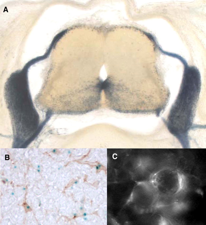
Characterization of cell populations expressing the wmN2 construct. A, Cross section, E14.5 embryo, demonstrating intense β-galactosidase histochemical labeling in spinal roots, DRGs, peripheral nerves, and the ventral portion of the neural tube. B, Neural tube cross section colabeled for β-galactosidase (histochemistry) and PDGFRα (immunohistochemistry) contains multiple colabeled cells, confirming that some transgene-expressing cells are within the oligodendrocyte lineage. C, Whole-mount preparation of a dorsal root ganglia from a 4-month-old wmN2 male chimera examined by fluorescence microscopy for eGFPlacZ reporter gene expression. Fluorescing cells, closely apposed to the large cell bodies of DRG neurons, are characteristic of DRG satellite cells.