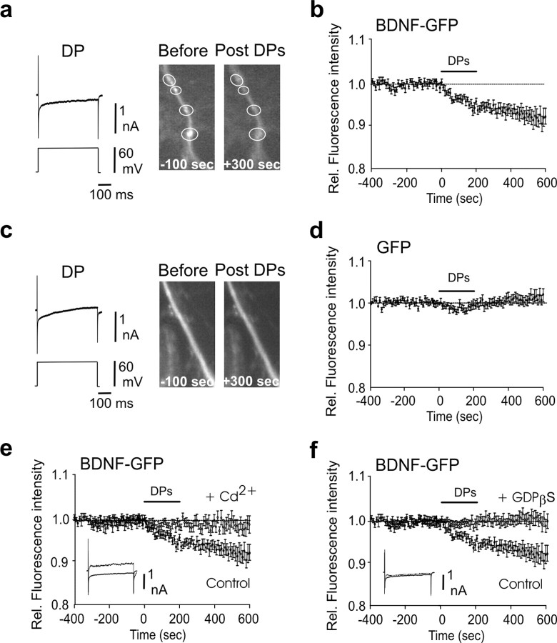
Figure 2.
Depolarizing steps trigger dendritic secretion of BDNF-GFP. a, Decrease of fluorescence intensity from BDNF-GFP granules localized in the dendrites was produced by 20 depolarizing steps of 50–60 mV depolarization, 500 ms long, given at 0.01 Hz. Note that fluorescence decreased within the white circle and did not increase in the surrounding area, indicating that the decrease in fluorescence intensity was not attributable to lateral movements in the x–y-axes. Left, Representative traces of a DP; note the inward Ca2+ current produced by the depolarization. Right, Example of dendritic BDNF-GFP granules (indicated by circles) before and after the DPs. b, Average time course of dendritic fluorescence change evoked by the DPs (n = 11). c, d, DP failed to produce significant variation of fluorescence in GFP-only-transfected neurons (n = 9). e, The effect of DPs on dendritic fluorescence was abolished by bath-applied Cd2+ (200 μm; n = 8); note the absence of Ca2+ current in response to DP (superimposed control and Cd2+ traces). f, Postsynaptic loading of GDP β-S (0.6 mm), a G-protein inhibitor that blocks granular secretion, prevented the DP-induced decrease of fluorescence in the dendrites of BDNF-GFP-transfected cells (n = 8); note that the Ca2+ current in response to DP was not affected (superimposed control and Cd2+ traces). All the experiments were performed in the presence of NBQX and APV. Rel., Relative.

