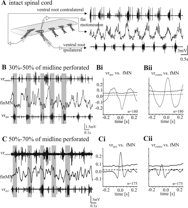
Figure 2.
Fin motoneuron activity during NMDA-induced fictive swimming. A–C, Recording of the membrane potential oscillation of a fin motoneuron together with the ipsilateral and contralateral ventral root in an intact spinal cord (A) and with intermediate midline lesions (B, C). In all experimental situations, an alternating activity pattern of the left and right side is evoked by superfusion of 150 μm NMDA. A, Recording of the membrane potential oscillation of a fin motoneuron in an intact spinal cord. Fin motoneurons show a clear out-of-phase activity with the ventral root discharge of the same side. B, When the midline of the spinal cord is lesioned 30–50%, clear out-of-phase activity of fin motoneurons with the ipsilateral ventral root activity persists. Bi, The mean waveform correlation of the cell shown in B with the rectified ipsilateral ventral root activity shows a clear negative peak around zero and therefore an out-of-phase activity. Here and in all subsequent figures, the dotted horizontal line close to zero denotes the SE, whereas the solid horizontal line denotes four times SE (see Materials and Methods). Bii, The mean waveform correlation with the contralateral ventral root discharge shows a clear positive peak around zero and therefore an in phase activity of the fin motoneuron. C, When the midline of the spinal cord is perforated 50–70%, fin motoneurons lose their out-of-phase activity and show instead an in-phase activity with the ipsilateral ventral root burst. The mean waveform correlation of the intracellular recording of the same cell with the rectified ventral root recording of the ipsilateral side shows a positive peak at zero (Ci) and no or a very weak negative correlation with the contralateral side (Cii).

