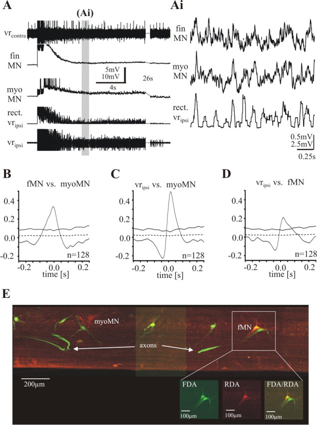
Figure 4.

Simultaneous recordings of a fin motoneuron and a myotomal motoneuron during electrically evoked motor burst activity in a piece of spinal cord lesioned 30–50% and electrically stimulated on the ipsilateral side. A, One sequence of a bout of activity. Top trace, Activity of the contralateral ventral root (vrcontra); second and third traces, intracellular recordings from a fMN and a mMN; fourth trace, rectified ipsilateral ventral root recording; fifth trace, activity of the ipsilateral ventral root (vripsi). Toward the end, 26 s are cut out (stippled lines) to increase the region of presentation. The gray bar indicates the episode of the recording shown in Ai in a higher time resolution without top and bottom trace. The fin motoneuron as well as the myotomal motoneuron receives excitatory synaptic input in-phase with the activity of the ipsilateral ventral root. B, Waveform correlations between the two motoneurons show a positive peak around zero. C, D, Waveform correlations of each motoneuron with the rectified ipsilateral ventral root recording show positive peaks around zero. E, Recorded motoneurons were filled with RDA (red marker) via the electrode. Fin motoneurons were retrogradely prelabeled with FDA (green marker). The yellow cell (fMN) is a double-labeled fin motoneuron (see inset).
