Figure 6.
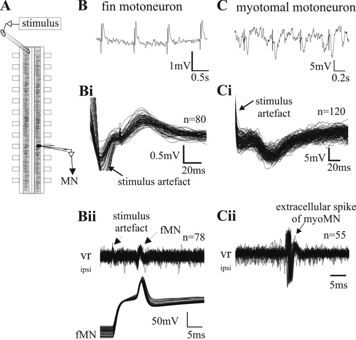
Connectivity of commissural long-range projecting interneurons to fin and myotomal motoneurons. A, Schematic drawing of the experimental set up for the stimulation experiments. The stimulus electrode is placed contralaterally eight segments rostral to the intracellular recording site. B, A stimulus of 2 ms duration leads to a compound EPSP in the fin motoneuron (tested between 0.001 and 0.03 mA stimulation strength). Bi, Multiple sweeps of the intracellular recording triggered by the stimulus artifact demonstrates the short, constant latency occurrence of the evoked EPSP, indicating a monosynaptic connection between projecting axons and fMN. Bii, During the experiment, a recorded neuron was identified as a motoneuron by injecting current into the cell to evoke orthodromic spikes, which were detected in the ventral root recording. To confirm the recording of a fin motoneuron, a dye was injected into the cell at the end of the experiment (data not shown). C, The same stimulus strength as used in B (tested between 0.001 and 0.03mA stimulation strength) at the same stimulus site evokes IPSPs in myotomal motoneurons. Ci, Multiple sweeps of the intracellular recording triggered by the stimulus artifact demonstrates the short and constant latency of the evoked IPSP between projecting axons and mMN. Cii, Identification of the cell shown in Ci as a motoneuron by the release of an action potential after rebound from inhibition with stronger rostral stimulation of the commissural interneurons. To confirm the recording of a myotomal motoneuron, dye was injected into the cell at the end of the experiment (data not shown).

