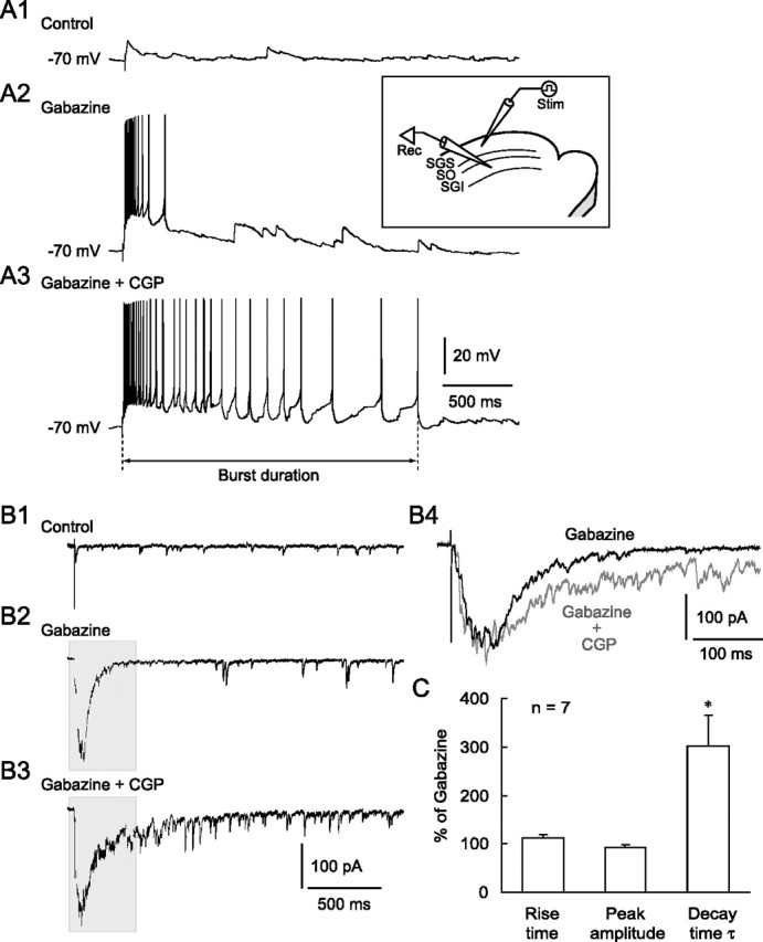
Figure 1.

Blockade of GABABRs prolonged the duration of burst activity. A1, SGS stimulation evoked EPSPs under control conditions in an SGI non-GABAergic neuron. Inset, Schematic showing the electrode arrangement. A stimulating (Stim) electrode was placed in the SGS. Whole-cell recordings (Rec) were obtained from SGI non-GABAergic neurons. A2, The SGS stimulation evoked bursts in this neuron after bath application of the GABAAR antagonist gabazine (10 μm). A3, Additional application of the GABABR antagonist CGP (3 μm) greatly prolonged burst duration, defined as the period between stimulation onset and the peak of the last spike. B1, Voltage-clamp recordings from the same neuron shown in A revealed that SGS stimulation evoked EPSCs in the control. Membrane potential was held at −60 mV. B2, Bath application of gabazine induced a burst of EPSCs in response to SGS stimulation. B3, Additional application of CGP increased the duration, but not the amplitude, of the EPSC bursts. B4, Expanded traces from shaded areas in B2 and B3 showed a similar peak amplitude but a prolonged decay time of the EPSC bursts when CGP was added. C, Summary graph showing that the rise time and peak amplitude of the EPSC bursts were not significantly changed, whereas the decay time constant (τ) was significantly prolonged by CGP. *p < 0.05, compared with gabazine alone (paired t test).
