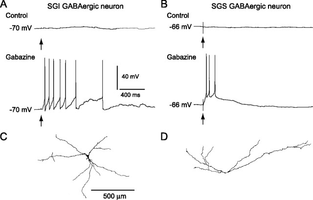
Figure 3.
Bursts of GABAergic neurons in the SGI and SGS. A, B, Under control conditions no clear responses were evoked in either SGI (A) or SGS (B) GABAergic neurons by SGS stimulation (top traces), whereas in the presence of gabazine (10 μm), the stimulation evoked bursts in both types of neurons (bottom traces). Arrows indicate stimulus timing. C, D, Somatodendritic morphology of the biocytin-filled SGI (C) and SGS (D) GABAergic neurons shown in A and B, respectively. Scale bar in C applies to D.

