
Figure 3.
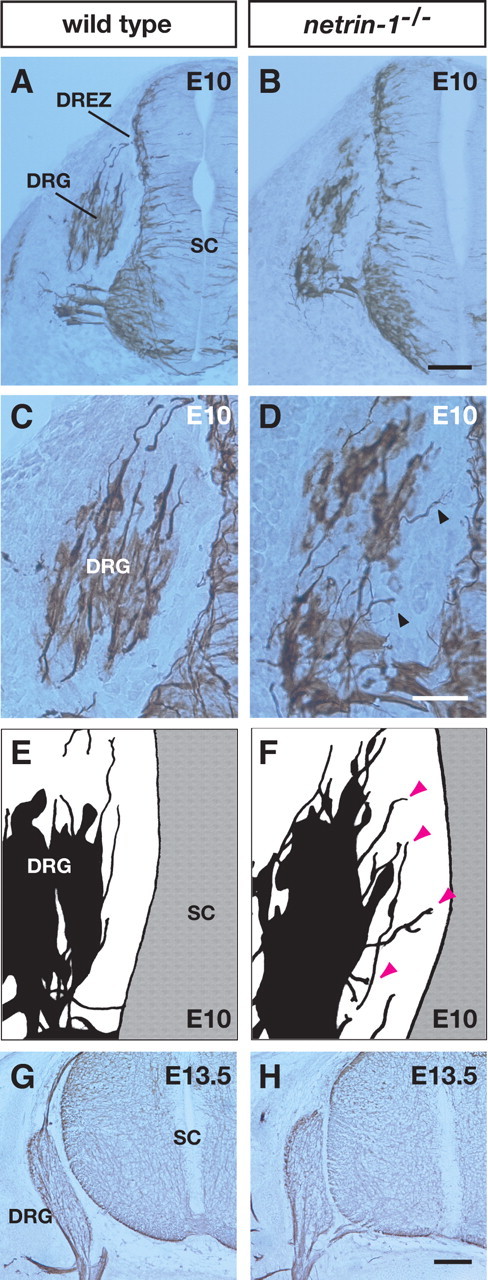
Defects in axonal trajectories at initial stages of DRG axonal projection in netrin-1 mutants. A,B, Transverse sections of E10 wild-type (A) and netrin-1 mutant (B) littermates labeled with an anti-class III β-tubulin antibody. C,D, Higher-magnification views of the DRG from A and B, respectively. Misrouted DRG axons are observed in netrin-1 mutant littermates (arrowheads). E, F, Camera lucida drawings showing axons and cell bodies of DRGs in sections of wild-type (E) and netrin-1 mutant (F) littermates. Arrowheads (magenta) indicate misrouted axons in the netrin-1 mutant. G, H, In contrast to E10, there are no aberrant DRG axonal projections in sections of E13.5 wild-type (G) or netrin-1 mutant (H) littermates. SC, Spinal cord. Scale bars: B, H, 100 μm; D, 50 μm.