Figure 7.
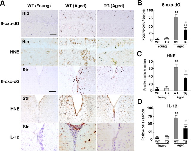
Amelioration of ROS-mediated oxidation of cell components and inflammation in the hippocampus of the aged TG mice. A, Immunohistochemical staining for 8-oxo-dG and HNE in the CA1 hippocampal subfield (Hip) and the periventricular area of the striatum (Str) of the young WT, the aged WT, and the aged TG mice. Scale bars, 50 μm. B–D, The mean number of positive cells for 8-oxo-dG (B), HNE (C), and IL-1β/cells (D) in the hippocampal CA1 subfield of the WT, the TG, and the LPS-treated TG mice of the aged group. Each column and bar represent the mean ± SEM of nine sections from three animals. The asterisks indicate significant differences versus the young group (**p < 0.01). The daggers indicate significant differences versus the aged WT mice (††p < 0.01).

