Figure 8.
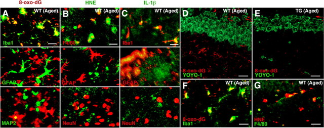
Immunofluorescent CLSM images for 8-oxo-dG, HNE, and IL-1β in the striatum and the hippocampus. A–C, Immunofluorescence for 8-oxo-dG (A), HNE (B), and IL-1β (C) with cell type markers (Iba1, F4/80 for microglia, GFAP for astrocytes, MAP2 and NeuN for neurons) in the periventricular area of the striatum of the aged WT mice. Scale bars: A–C, 30 μm. D, E, Immunofluorescence for 8-oxo-dG (red) with YOYO-1-stained neurons (green) in the hippocampal CA1 subfield of the aged WT (D) and TG (E) mice. Scale bars: D, E, 20 μm. F, G, Immunofluorescence for 8-oxo-dG (F) and HNE (G) with markers of microglia (Iba1 and F4/80) in the hippocampal CA1 subfield of the aged WT mice. Scale bars: F, G, 5 μm.

