
Figure 3.
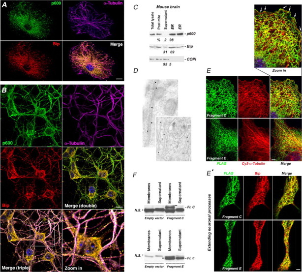
p600 associates with the ER. A, B, Colocalization of p600 with MTs and the ER marker Bip in nondifferentiated CAD cells (A) and primary mouse cortical neurons (B) in a vesicular/punctate pattern. C, Crude microsome ER preparation from adult mouse brains. Post-mito, Postmitochondrial fraction containing ER microsomes before purification; supernatant, fraction discarded before collection of microsomes. The amount of each protein in each fraction is expressed in percentage. D, Immunogold labeling of ER tubules with p600 antibodies. E, Polygonal reticular pattern of peripheral ER tubules exhibited by the fragments C and E in nondifferentiated CAD cells. These ER tubules are juxtaposing MTs up to the cell edge (white arrows). E′, Colocalization of FLAG-tagged fragment C or E with the ER marker Bip in differentiated CAD cells. F, Fragment C (Fr. C) (double black stars) is enriched in the membrane fraction, whereas fragment E (Fr. E) (double black stars) is found in both membrane and nonmembrane fractions. N.S. refers to nonspecific bands. Cells transfected with an empty vector were used as control. All pictures were taken with a confocal microscope. Scale bars: A, E′, 25 μm; B, E, 10 μm; D, 200 nm.