
Figure 4.
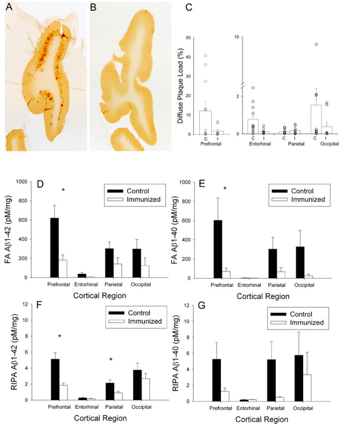
A, B, Diffuse prefrontal Aβ plaques were extensive in a control animal (A) and significantly reduced in an immunized animal (B). C, Animals chosen for this illustration had Aβ plaque loads similar to the group means of the control and immunized animals. Consistent with immunolabeling for Aβ, diffuse plaque load was reduced in the prefrontal cortex, the entorhinal cortex, and the occipital cortex but not in the parietal cortex. Each symbol represents an individual animal. D, E, Formic acid-extracted Aβ1–42 (D) and Aβ1–40 (E) were both significantly reduced in immunized dogs in multiple cortical regions. F, G, Similarly, RIPA-extracted Aβ1–42 (F) and Aβ1–40 (G) were reduced in treated dogs. Bars represent means, and error bars represent SEM. *p < 0.05.