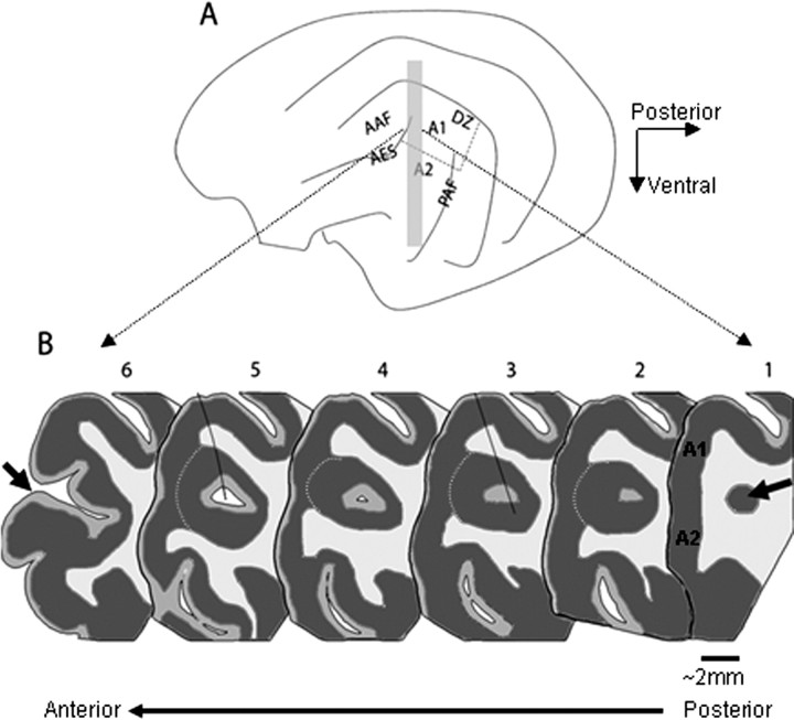
Figure 1.
Anatomy of the anterior ectosylvian sulcus. A, Schematic diagram depicting the lateral view of the cat's left hemisphere, including the main cortical auditory areas: A1, A2, the anterior auditory field (AAF), the PAF, the DZ and the AES. B, Schematic representation of six coronal sections from different locations along the longitudinal axis of the AES (the approximate location of these sections is indicated by the gray rectangle in A). The arrow in section 1 indicates the posterior pole of the AES, which consists of a small cluster of cells that increases in size more rostrally, and which lies underneath the auditory fields of A1 and A2. Dark gray corresponds to gray matter, and white corresponds to white matter, whereas intermediate gray represents layer I, a region poor in cells that appears in the middle of the cell cluster forming the posterior pole of AES. The pAES is defined as the part of AES that lies buried underneath the middle ectosylvian gyrus (sections 1–5), and aAES is the open part of the sulcus (represented here by section 6, with the sulcus lips anterior to the opening depicted by an arrow). The white dashed line on sections 2–5 is indicating layer 5, the border between the gray matter of AES and that of A1 and A2. The black lines that partially cross sections 3 and 5 indicate two electrode penetrations that were reconstructed in this brain.

