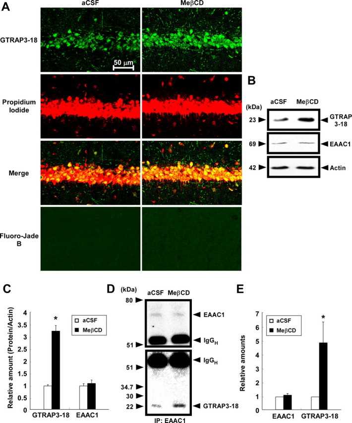
Figure 6.

Effects of MeβCD on GTRAP3-18 expression and GSH contents in vivo. Adult male C57BL/6J mice (n ≥ 3) were treated intracerebroventricularly for 5 d with either MeβCD (40 mg/ml; flow rate, 0.5 μl/h) or aCSF. A, GTRAP3-18 immunolabeling (green), propidium iodide counterstaining (red), and Fluoro-Jade B staining (green) were performed in the CA1 field of the mouse hippocampus and examined by confocal laser-scanning fluorescence microscopy. B, Immunoblot analysis of the mouse hippocampus was performed using each specific antibody. C, Protein bands were quantified against the amounts of actin in the same lysates. D, After immunoprecipitation using lysates of mouse hippocampus and anti-EAAC1 antibody, immunoblot analysis was performed. E, GTRAP3-18 levels were quantified against EAAC1 amounts in the precipitates. F, GSH contents of the mouse hippocampus were measured by the NADPH-dependent GSH reductase method. *p < 0.05 compared with aCSF.
