
Figure 4.
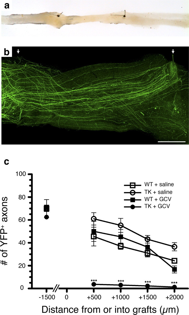
Regeneration of peripheral axons is prevented in the absence of CD11b+ cells. a, Photograph showing an example of sciatic–sciatic nerve graft. b, Representative fluorescence photomicrograph showing sciatic nerve axons expressing the YFP marker that have regenerated into a PN graft collected from a saline-treated YFP−/TK+ mouse (i.e., CD11b-TKmt-30 +/−). The arrows point to the two 10-0 sutures used to connect the PN graft between the proximal and distal ends of the recipient sciatic nerve from the YFP transgenic mouse. c, Quantification of the number of YFP-labeled axons at predetermined distances from the proximal host–graft interface (n = 8 per group). Although the total number of YFP+ axons was similar in all groups at a distance of 1.5 mm from the proximal host–graft interface (i.e., into the proximal end of the recipient sciatic nerve), YFP+ axons were only able to regenerate into predegenerated PN grafts in the presence of CD11b+ cells. Error bars indicate SEM. ***p < 0.001. Scale bar, 500 μm.