
Figure 9.
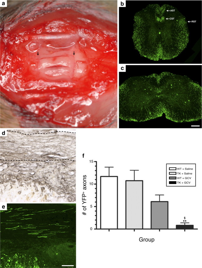
Regeneration of SCI axons into PN grafts is prevented in the absence of CD11b+ cells. a, Photograph showing three sciatic nerve segments transplanted into the spinal cord of a T12/L1 dorsal hemisected Thy1-YFP-H+/− transgenic mouse. The arrows point to PN grafts/spinal cord tissue interfaces. b, c, YFP expression in the thoracic (b) and lumbar (c) spinal cord of a Thy1-YFP-H+/− transgenic mouse. Note the presence of fluorescence in axons traveling through most, if not all, spinal cord projection systems, including the descending corticospinal (CST) and rubrospinal (RST) tracts and the ascending sensory tract (AST). d, e, Visualization of laminin immunolabeling (d) (to visualize PN tissue) and YFP fluorescence (e) on adjacent spinal cord sections revealed that PN grafts are densely penetrated by regenerating SCI axons at 2 weeks after SCI/grafting. The dotted lines in d indicate the anatomical boundaries of a PN graft. f, Quantification of the number of YFP-labeled axons at predetermined distances from the rostrocaudal host–graft interface. Note that SCI YFP-labeled axons did not regenerate into predegenerated PN grafts lacking CD11b+ cells. Error bars indicate SEM. **p < 0.01 compared with the WT plus saline group; § p < 0.05 compared with the TK plus saline group. Scale bars: (in c) b, c, 275 μm; (in e) d, e, 100 μm.