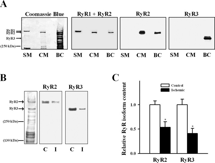
Figure 6.
Presence of different RyR isoforms in ER vesicles. A, Proteins bands of SR from skeletal (SM) and cardiac (CM) muscle and ER from rat brain cortex (BC) were separated by gel electrophoresis and electrotransferred to a PVDF membrane (for details, see Materials and Methods). Reactivity to antibodies that recognize RyR1 together with RyR2 (RyR1 + RyR2), RyR2, or RyR3 are shown. After exposure to the different antibodies, the membrane was stained with Coomassie blue to reveal protein bands (first left blot). B, Reactivity of ER vesicles isolated from control (C) and ischemic (I) brain cortex to specific RyR2 or RyR3 antibodies. A Coomassie blue-stained membrane for control ER vesicles is shown on the left side. C, The RyR2 and RyR3 contents in ER of control and ischemic brains were estimated from Western blot analysis; values were normalized by their respective controls. *p < 0.05, content differed from the corresponding control.

