
Figure 5.
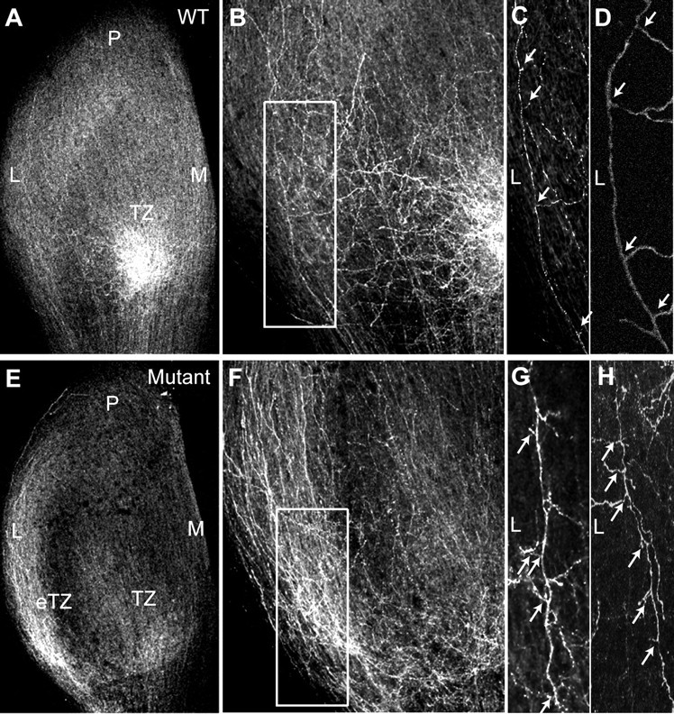
Abnormal directional branch extension in WT and L1(Y1229H) mutant mice at P2. A, B, Labeling of RGC axons at P2 after focal injections of DiI in the ventrotemporal retina of WT mice show interstitial branches extending toward the nascent TZ in the anteromedial SC. C, D, Most of the branches extending from axons that navigate lateral to the future TZ are oriented medially as shown in a higher magnification of the boxed area in B from confocal z-stacks (arrows). E, F, DiI labeling of VT axons in the SC of L1(Y1229H) mice at P2 reveal an accumulation at an inappropriate ectopic TZ (eTZ) as well as at a more appropriate future TZ. G, H, Many interstitial branches of mutant VT axons are abnormally oriented toward the lateral SC, as shown in a higher magnification of the boxed area in F from z-stacks (arrows). M, Medial; L, lateral; P, posterior.