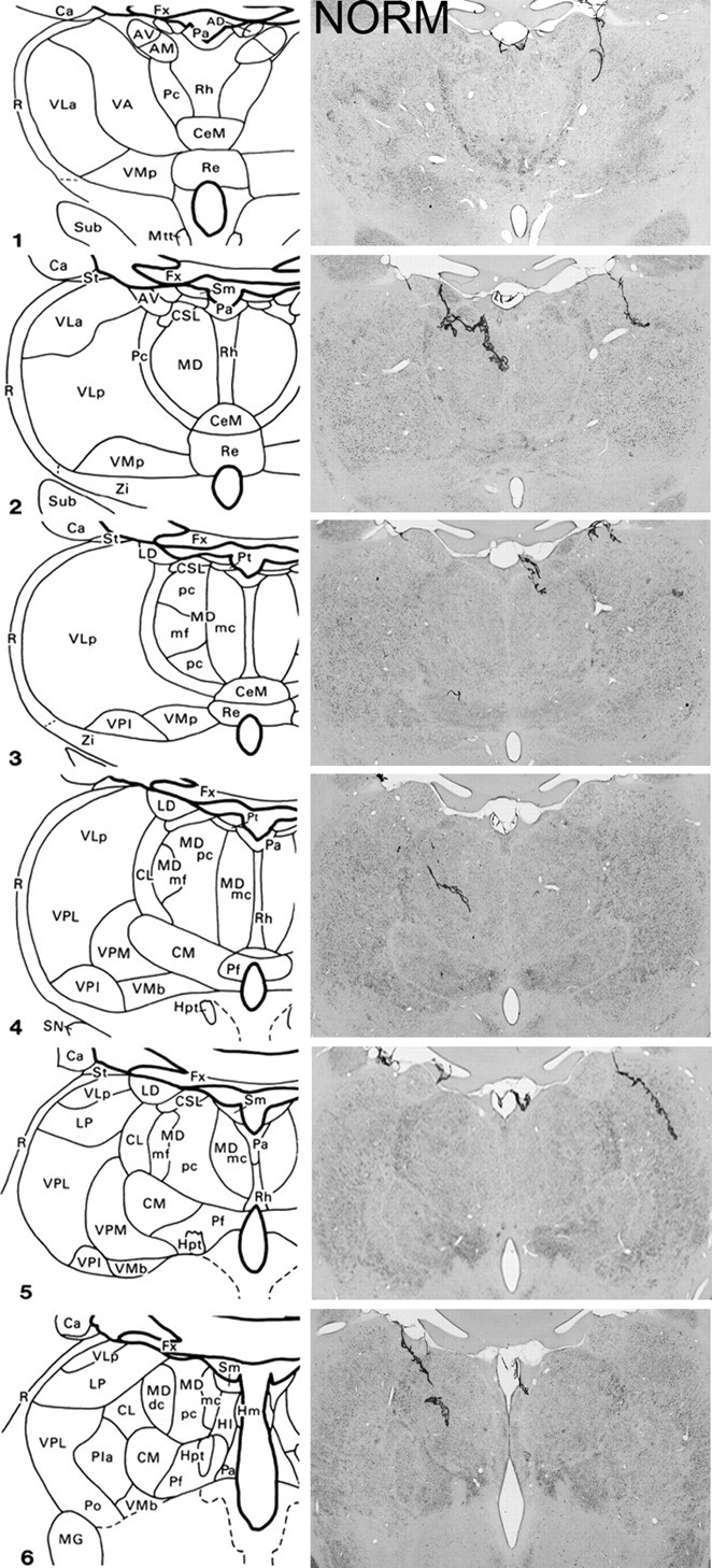
Figure 1.

Schematic diagrams of six sections, 1 mm apart, through the medial thalamus of a monkey taken from Gaffan and Murray (1990) and photomicrographs of a normal medial thalamus (NORM) corresponding as closely as possible to the schematic diagrams. For abbreviations, see Mitchell et al., (2007a).
