
Figure 1.
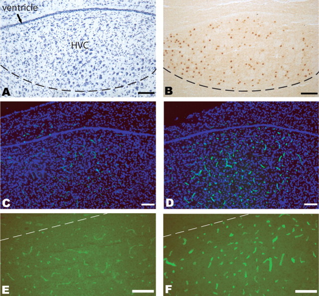
Testosterone induces MMP activity within the HVC microvasculature. A, Nissl-stained HVC section of adult female canary brain, including HVC at the dorsal surface of the mesopallium. For terminology used, see Reiner et al. (2004) and Jarvis et al. (2005). B, Estrogen receptor-α immunoreactivity was used to define the borders of HVC for cell quantification purposes. C, D, In situ zymography using Oregon 488-conjugated gelatin was used to localize gelatinase activity (green), in sections counterstained with DAPI (blue). C, A low-magnification image of HVC and its surround in an unimplanted adult female canary. D, A matched adult female experimental given testosterone 9 d before being killed. High signal in the dorsocaudal mesopallium is evident, and essentially restricted to HVC. E, F, Higher-magnification images, without counterstaining, of zymography-defined HVC MMP activity in an unimplanted control (E) and in a testosterone-implanted bird (F). Scale bars, 100 μm.