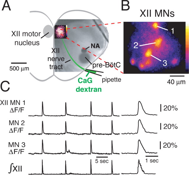
Figure 3.
Functional imaging of CaG-labeled inspiratory XII motoneurons. A, Rhythmically active XII MNs were labeled retrogradely by CaG-dextran loaded over 6–8 h through a suction electrode applied to the cut end of the rostralmost XII nerve root. Localized inspiratory population CaG fluorescence “flash” image in the XII motor nucleus is shown superimposed on IR-DIC image of the slice. B, High-magnification (63× composite), background-subtracted, and pseudocolored flash image shows individual inspiratory MNs imaged simultaneously (7 neurons shown) in a single focal plane. C, Rhythmic elevations in CaG fluorescence (ΔF/F) in three inspiratory MNs (neuron somata labeled 1–3 in B) in phase with inspiratory XII nerve discharge (∫XII); expanded time base of CaG fluorescence transients during inspiratory burst are shown at right. NA, Semicompact division of nucleus ambiguus.

