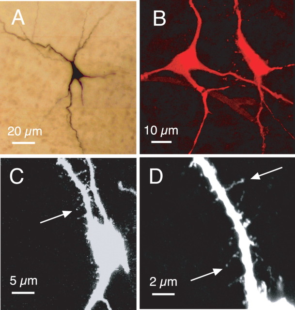
Figure 8.
Morphological features of somata and primary dendrites of pre-BötC inspiratory neurons. A, Light microscopic image (composited from multiple merged 20× magnification photomicrographs) of intrinsic burster inspiratory neuron in situ in whole-mounted fixed slice preparation. B, Confocal image of two nonintrinsically bursting pre-BötC neurons labeled with Texas Red (100×; two-dimensional projection from merged image stack; 615 nm emission wavelength). C, D, Confocal images (63× magnification in C; 100× in D) of Texas Red-labeled nonintrinsically bursting pre-BötC inspiratory neuron, showing examples of spines on primary dendrites. Image in C, showing several primary dendrites, is a two-dimensional projection from the merged image stack. Higher-magnification image in D is focused on a single primary dendrite of the cell shown in C (note that the medial dendrite in C is out of the focal plane and therefore does not appear in the image shown in D). Original color projection images have been inverted into black-and-white for clearer resolution of dendritic spines.

