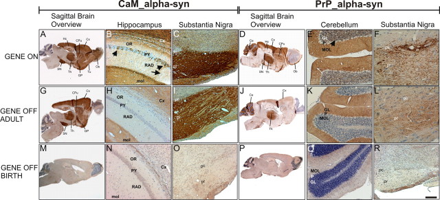
Figure 2.
Distribution of human α-syn expression in treated and untreated conditional tg mice. Sagittal sections showed regional immunostaining for human α-syn in the brain of adult untreated CaM_α-syn mice (A–C) and PrP_α-syn mice respectively (D–F). A, Strong immunoreactivity was detected in the olfactory bulb (Ob), olfactory tubercle (Tu), cortex (Cx), caudate–putamen (CPu), globus pallidus (GP), substantia nigra (SN), thalamus (Th), and hippocampus (Hc) in CaM_α-syn mice. B, In the CA1 area of the Hc, α-syn immunoreactivity was observed in a subpopulation of the pyramidal cells (PY) (arrows), various dendrites in the stratum radiatum (RAD) (arrowhead), and in the neuropil of the molecular layer (mol). C, Intense α-syn staining was observed in both the substantia nigra pars compacta (pc) and pars reticulata (pr). D, PrP_α-syn mice displayed minor expression in the Ob, the CPu, the GP, the Hc, and the pr in comparison with CaM_α-syn mice. Additional staining was observed in the cerebellum (Ce). E, Expression of α-syn was detected in the molecular layer (MOL) and in the granular layer (GL), without staining of the Purkinje cell layer (P). F, Staining was prominent in the pc but less expressed in the pr. Doxycycline treatment over a 3 week period of adult CaM_α-syn mice (G–I) and adult PrP_α-syn mice (J–L) partly ceased transgene expression. H, I, In CaM_α-syn mice, treatment resulted in complete loss of immunoreactivity in the stratum oriens (OR), PY, and RAD of Hc, whereas the mol, the overlying Cx (H), and neuronal processes of the SN (I) were still stained, although less prominent. J–L, In treated PrP_α-syn mice, residual α-syn expression was detected in the Th (J) but abolished in the GL (not in the MOL) of the Ce (K) and reduced in the pc and pr of the SN (L). α-Syn staining was missing in CaM_α-syn mice (M–O) and PrP_α-syn mice (P–R), when mice were born and raised (over a minimum of 8 weeks) with dox. Scale bar: B, C, E, H, I, K, N, O, Q, R, 20 μm; A, D, G, J, M, P, 200 μm; F, L, 5 μm.

