Figure 7.
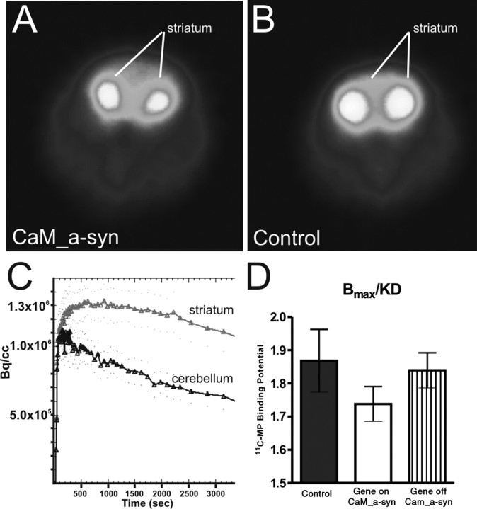
Striatal DAT binding potential quantification in conditional mice. A, B, Representative transverse color-coded microPET images (of all frames over 60 min acquisition time) of striata were conducted with [11C]d-threo-methylphenidate of a CaM_α-syn (A) and the respective control mouse (B). C, Time–activity curve of striatum and cerebellum of a CaM_α-syn mouse were normalized to the injected dose. TACs from striatum and cerebellum were well separated, indicating that the injected tracer did not cause saturation effects or unspecific binding. D, Quantitative analysis of DAT binding potential in the striatum of CaM_α-syn mice (n = 5), respective controls (n = 5), and dox-treated CaM_α-syn mice (n = 3; gene off) revealed reduced DAT binding without reaching statistical significance in CaM_α-syn mice. Error bars indicate SEM.

