
Figure 2.
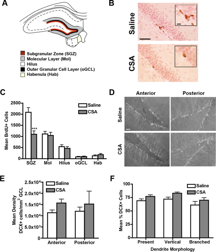
CSA decreases proliferation in the SGZ, but does not alter DCX+ neuron number or dendritic morphology. A, Schematic of the four regions of the dentate gyrus and medial habenula in which cell counts were collected for this study. B, Representative images at a proliferation time point (2 h post BrdU) injection show the decreased number of BrdU+ cells in the SGZ after CSA. BrdU+ cells (brown, stained via DAB IHC) appear in the SGZ on the border of the granule cell layer labeled (pink, stained with Fast red) and the hilus. Inset is a magnification of the BrdU+ cell clusters. Scale bar, 50 μm; inset, 10 μm. C, Quantitative analysis of BrdU+ cells in the dentate gyrus after CSA found a decrease in proliferation specific to the SGZ. D, Representative images of DCX+ cells in the SGZ at an anterior section (−2.80 mm from bregma) and posterior section (−6.60 mm from bregma) show staining of both the cell bodies and processes in both saline and CSA rats. Scale bar, 50 μm. E, Quantitative analysis of density of DCX+ cells in the SGZ after CSA found no change. F, CSA did not change the dendritic morphology of DCX+ cells in the posterior SGZ. Data for C, E, and F are presented as mean ± SEM. Data in C multiplied by fraction of sections examined to result in BrdU+ cell number. ***p < 0.001.