Figure 1.

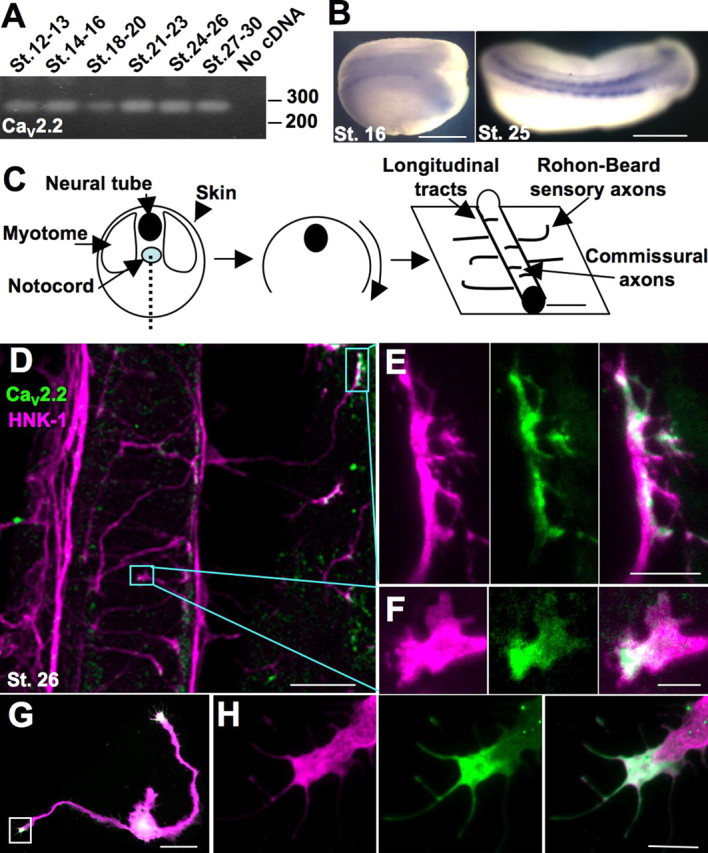
CaV2.2 is expressed in the developing Xenopus neural tube and is localized to the growth cone during axon outgrowth in vivo and in vitro. A, RT-PCR of a 272 base pair fragment of CaV2.2 using mRNA collected from dissections of dorsal halves of embryos from stages (St.) 12–30. B, In situ hybridization using the probe made from the same fragment reveals expression of CaV2.2 in the developing neural tube. Scale bar, 500 μm. C, Schematized dissection for immunostaining: a ventral incision was made to remove the myotomes and notochord and reveal the neural tube and skin. Staining with the neuronal marker HNK-1 reveals longitudinal tracts running anteroposteriorly, axons of sensory Rohon-Beard neurons innervating the skin, and commissural interneurons crossing the ventral midline. D, Ventral view of a spinal cord and skin of a dissected embryo stained for HNK-1 (purple) and CaV2.2 (green). Boxes outline the CaV2.2-positive growth cones pictured in E and F. Scale bar, 50 μm. E, Sensory Rohon-Beard growth cone expressing CaV2.2 (green). Scale bar, 10 μm. F, Commissural interneuron expressing CaV2.2 (green). Scale bar, 5 μm. G, Cultured neuron stained for β-tubulin (purple) and CaV2.2 (green) expressing CaV2.2 in the growth cone. Scale bar, 50 μm. H, Growth cone boxed in G. Scale bar, 10 μm.
